
Let us discuss the posterior arm chain and body movements that address the area . . . . . . . . . .
In kinesiology, the posterior arm chain refers to the muscles located on the back of the arm, primarily including the triceps brachi. This chain of muscles works to extend and retract the arms and trunk, stabilizing the shoulder joint and plays a crucial role in various movements, from pulling to throwing.
The concerned movements address extension / abduction / adduction of the arms, extension of the fingers, and stabilization of the shoulder blades. In modernized society, many people do not partake in activities that maintain strength and function of these musculatures. Decreased muscle strength in these areas leads to compromises in posture, breathing and shoulder functioning.
Description
Actions
Exercises
Description
The posterior arm chain is crucial to stabilizing the shoulders, maintaining posture and facilitating better breathing. Movements of extension, abduction, adduction are performed as designed when these musculatures function correctly. These muscuIatures, when working in synchronization, initiate or stabilize many movements that are associated with the upper body. Posture would be un-manageable in the absence of the muscle groups. These muscles are key to upright posture; they help to withstand the ever present effects of gravity. The necessity of breathing cannot be overstated, as breath is life. The muscles can be engaged such that the upper chest can be more open and receptive to greater oxygen uptake.
The shoulders are an important joint. They transfer forces from the lower body, to the upper body, and out through the arms. The shoulder is the most mobile joint in the body. The muscles that are associated with it must work in unison to support the optimal functioning in this area Reaching overhead, out to the side, forward, rearward . . . are all dependent on shoulder stability / mobility.. Both of these qualities require that shoulder blades and associated musculatures function as seamlessly as possible.
The societal trend for forward head orientation serves to compromise the performance of the PAC muscles. This ‘trend’ (of forward head) causes lengthening of the shoulder blade retractors and upper traps along with associated neck musculatures. This can lead to rounded shoulders which can tightening muscles in the front of the body. Sitting down to drive to and fro, sitting in front of the television, using electric bicycles . . . these modern day conveniences reduce the amount of time that people beneficially engage the body. utilizing proper posture which will support patterns of good muscular functioning and alignment.
Most of us use what breathing expert Dr. Belisa Vranich calls “vertical breathing.” When you breathe vertically, your shoulders go up during the inhale and everything in your torso stretches up vertically. When you exhale, your shoulders go back down. But vertical breathing doesn’t just diminish our inhaling; it also messes up our exhaling. A vertical breath does not utilize the diaphragm, or the associated antagonist muscles (of exhalation), as designed. The muscles of the posterior (arm) chain are designed to supplement the breath. As Dr. Vranich explains, by not fully exhaling, you don’t allow your lungs room to breathe in fresh, new air.
To maintain functionality of over a lifetime requires that structural challenges placed upon the body be addressed. Movements that can contribute to functionality of the posterior arm chain are basic. They do not build strength as much as they assist the body in maintaining function by developing the deeper musculatures that keep the body doing, wherever that may entail.
Actions
According to the Modern Art and Science of Mobility (Broussal-Derval, Ganneau) the superficial ‘surface’ muscles are designated as:
- Upper trapezius
- Middle trapezius
- Lower trapezius
Upper Trapezius
The upper trapezius, the part that goes across the tops of your shoulders, can elevate or bring up your shoulder girdle. It also helps extend, tilt, and rotate your neck, which has the effect of bringing your head back, to the side, and turning it. The rotation function takes the head to the opposite side of where this neck and shoulder muscle is located.
Together with the lower traps, the upper trapezius also helps rotate your shoulder blade upward. This movement occurs when you lift your arm up to the side, providing your shoulders, neck, and upper back are in good alignment and your muscles are flexible.
Middle Trapezius
The middle trapezius helps bring the shoulder blades back, toward the spine. Again, if you sit at a desk or drive all day, this may prove a handy move for preventing or managing excessive kyphotic (rounding of the upper back) posture in that area. The middle trapezius also helps stabilize the shoulder during certain arm movements.
Lower Trapezius
Finally, the lower trapezius muscle is tasked with the upper and mid-spine stabilizing action of bringing the shoulder girdle down. This is the opposite action to that of the upper trapezius.
The trapezius, as an accessory breathing muscle2,helps open up a small amount of breathing room in the upper chest area.
The main functions of the deltoid muscles are to stabilize the shoulder joint and move the humerus at or through the shoulder. These muscle movements are:2
- Abduction: Lifting your arm outward to the side of your body
- Flexion: Moving your arm forward and up over your head
- Extension: Moving your arm behind you
- Rotation: The humerus rotates in the socket of the shoulder.
The deltoid muscles have three heads, only the lateral and posterior heads are of concern for this blog post, that attach to bone and other tissue. Their origins—where the muscle attaches to a fixed point that does not move when the muscle contracts—are the clavicle and scapula. Their insertion—the point that attaches to a bone that moves—is the humerus.
The lateral deltoid, located in the middle of the shoulder, lifts the humerus to the side (laterally) away from the body. During abduction, the lateral deltoid moves downward to accommodate the arm’s outward movement.
The posterior deltoid, located at the back of the shoulder, also rotates the joint laterally to move the arm outward and backward. During extension, the posterior deltoid allows the humeral head to rotate downward and inward in the socket of the joint.
These tissues are in place to provide overall structure to the body proper. They form a secondary skeleton that provides for muscle attachment. These tissues enclose muscle fibers. They form fibrous networks that dissipate stress at points of connection. They are integral to force transfer between the radius, ulna and humerus. They contribute to the proper functioning of the shoulder musculature.
The extensor digitorum communis is a superficial extensor muscle located in the posterior compartment of the forearm. [1] It shares a common synovial tendon sheaths along with other extensor muscles which helps to reduce friction between the tendon and the surrounding structures. As it courses the dorsum of the hand, the extensor digitorum communis muscle spreads out into four (4) flat tendons deep to the extensor retinaculum to the medial four fingers.
Primarily, the extensor digitorum communis extends medial four digits at the metacarpophalangeal joints and secondarily at the interphalangeal joints. [1] It also acts to extend the wrist joint.
According to the Modern Art and Science of Mobility (Broussal-Derval, Ganneau) the deep ‘structural” muscles are designated as:
The rhomboids consist of two separate muscles; the major and minor muscles, which are found immediately deep to the trapezius. The rhomboid minor is a cylindrical muscle that originates at the ligamentum nuchae and C7 and T1 vertebra. It inserts at the scapula’s medial border near the base of the spine of the scapula. The rhomboid major is a quadrangular muscle located inferior to the rhomboid minor. The origin of the rhomboid muscles is from the spinous processes of the T2-T5 vertebra and inserts on the medial border of the scapula, just inferior to the rhomboid minor.[2][3][4]
With the assistance of many other muscles, the rhomboids help form the shoulder girdle. This group of muscles is important for the movement of the upper extremity and stabilization of the shoulder through articulation with the trunk. Functionally, the rhomboid muscles retract, elevate and rotate the scapula. They also protract the medial border of the scapula, keeping it in position at the posterior thoracic wall. Dysfunction, weakness, or loss of nerve function to the rhomboids causes winging of the medial border of the scapula and inferior scapular angle rotation.[2][5]
The rhomboids are also vital to actions such as pulling and have been shown to play a large role in throwing and overhead arm movement
Infraspinatus is a thick triangular muscle that occupies the majority of the dorsal surface of the scapula. It arises mostly from the infraspinous fossa of scapula, and connects it to the proximal humerus. Rotator cuff muscles form a functional musculotendinous unit that both stabilizes and produces movements on the shoulder joint. The function of this muscular apparatus is to provide a wide range of arm motion while keeping the head of humerus centralized within the glenoid cavity. Acting as a prime mover, infraspinatus is a powerful lateral rotator of the humerus. This action is important in preloading the upper extremity in backward extension and external rotation on the shoulder joint, seen in actions such as hitting overhead. The muscle is contracted eccentrically to smoothen the upper extremity during deceleration of powerful overhead movements
The triceps, or triceps brachii, is the large muscle found at the back of the upper arm. Along with the other muscles of the arms and legs, the triceps muscle is a voluntary muscle, meaning it’s a muscle you choose to move. The triceps are made up of skeletal muscle fibers that contract under conscious control. It is a fusiform, or spindle-shaped, muscle that is wider in the middle and narrower at each end.1
“Triceps” means “three heads,” which refers to the subdivision of the triceps muscle into three different segments called heads. These include the following:2
- Long head: Running from the shoulder blade down along the length of the humerus (upper arm bone)
- Lateral head: Located on the outer side of the back of the arm
- Medial head: Located on the inner side of the back of the arm
All skeletal muscles connect to bones via tendons. Each head of the triceps has a separate short tendon that attaches the muscle heads to the shoulder blade and arm bones where they originate. All three heads merge together and form one thicker and longer tendon that attaches the entire muscle to the elbow.
Though they might be easy to ignore, your triceps are an important element for helping your entire upper body functioning properly. Fun fact: The muscle actually makes up two-thirds of your entire arm. “The tricep muscles extend the shoulder and the elbow joint, and building up tricep strength, stability, and control can improve flexibility in addition to improving posture,” says Joey Cifelli, a master trainer at Crunch Gym in New York City
Improved Posture: Triceps exercises can help improve posture by strengthening the muscles that support the spine and shoulders.
The outer surface of the ulna (except at the joints) is covered in a thin fibrous layer known as the periosteum. Periosteum is made of a dense weave of collagen fibers that extend into the tendons and ligaments that attach the ulna to the muscles and bones of the arm.
The dorsal border starts posterior to the olecranon process and runs down to the styloid process as well. Here, it works as attachment sites for the flexor carpi ulnaris, extensor carpi ulnaris, and the flexor digitorum profundus. The interosseous crest starts at the radial notch and then ends at the ulnar head, giving attachment to the interosseous membrane, a compact fibrous area of connective tissue filling the space between the ulna and the radius.[4][5][6]
The dorsal surface consists of the posterior and the lateral sides of the bone and houses the insertion sites for the anconeus muscle and the supinator. However, more distally, it also is involved in attachments of the extensor carpi ulnaris, supinator, abductor pollicis longus, extensor pollicis longus, and the extensor indicis propius.
- Extensor carpi ulnaris – posterior border
- Supinator – proximal ulna
- Abductor pollicis longus – posterior surface
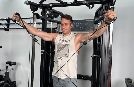
Exercises
Triceps extension using a suspension trainer

Expanded cross – use exercise bands to innervate the overhead press pathways – see video
Lateral Kettlebelll Swing

Pike push up

National Library of Medicine. Anatomy, shoulder and upper limb, deltoid muscle.
https://pmc.ncbi.nlm.nih.gov/articles/PMC4756000/
https://pmc.ncbi.nlm.nih.gov/articles/PMC6405356/
Landin D, Thompson M, Jackson M. Functions of the Triceps Brachii in Humans: A Review. J Clin Med Res. 2018 Apr;10(4):290-293. doi: 10.14740/jocmr3340w.
